Venue
Madrid´s Institute for Material Sciences (ICMM-CSIC), Madrid, Spain
Madrid, June 16 – 20, 2025
How to find us: Contact | ICMM
Overall Aims and Course Outline
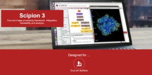
Cryo-Electron Microscopy (Cryo-EM) allows the determination of three-dimensional structures of biological macromolecules at atomic resolution. This course provides an overview of image processing in Single Particle Analysis (SPA) using Scipion. In the first part of the course, participants will learn the fundamentals of reconstructing biological structures from single particle images obtained through cryo-EM, employing state-of-the-art software in the field.
The second part of the course will focus on interpreting reconstructed maps through atomic modeling. We will use the Scipion platform, integrating tools such as Coot, Chimera, Refmac, and AlphaFold. This course is open to researchers of all levels.
The course will be held in-person and is designed for researchers at any stage of their career who are interested in incorporating Cryo-EM into their research.
Expected Impact for Young Researchers
This course will equip young researchers with the knowledge and skills necessary to apply Cryo-EM in the study of biological macromolecules. Through training in image processing and the interpretation of reconstructed maps, participants will be able to address the structural characterization of biomolecules. These approaches will enhance the understanding of molecular mechanisms and facilitate the use of Cryo-EM as a tool in biomedical and biotechnological research.
Tutors
Marcos Gragera(MG) – Single Particle Analysis specialist, BCU/I2PC
Marta Martinez (MM) – Modelling specialist, BCU/I2PC
Roberto Marabini (RM) – Professor, UAM, Collaborator BCU/I2PC
Deborah Cezar Mendonca – Biologie structurale intégrative (IGBMC)
Registration Fee
- Academic Registration Fee Instruct countries: Predocs & Postdosc 50€
- Academic Registration Fee Instruct countries: Others 100€
- Academic Registration Fee Non Instruct countries: Predocs & Postdosc 100€
- Academic Registration Fee Non Instruct countries: Others 150€
- Industrial Registration Fee 300€
Acommodations
- Hotel Chamartín The One (****): Calle Agustín de Foxá, s/n 28036 Chamartín, Madrid. Reservas: reservas@hotelchamartintheone.com
- Hotel Exe Plaza (****): Paseo de la Castellana 191, Madrid, 28045, España. Reservas: reservas@hotelexeplaza.com
- Hotel Crisol Via Castellana (****): Paseo de la Castellana, 220, Madrid, 28046, España. Reservas: reservas@hotelviacastellana.com
- VP Jardín de Tres Cantos (***): Avenida de los Encuartes, 17, Tres Cantos, 28760, España. tc@vphoteles.com
Additionally, on our campus, there is a student residence that might have rooms available for those dates. Here are the links:
Residencia UAM: https://resa.es/residencias/madrid/erasmo/
Closes on 23 May 2025 at 12:00 PM
|
Day 1 |
|
|
10:30 – 11:00 |
Badge Pick-up & Welcome |
|
11:00 – 13:00 |
Movie alignment, screening micrographs and CTF |
|
13:00 – 14:00 |
Lunch Break |
|
14:00 – 16:00 |
Particle picking and screening |
|
16:00 – 16:30 |
Coffee break |
|
16:30 – 17:30 |
2D classification |
|
17:30 |
End of day |
|
Day 2 |
|
|
09:30 – 11:00 |
Initial volume |
|
11:00 – 11:30 |
Coffee break |
|
11:30 – 13:00 |
3D classification |
|
13:00 – 14:00 |
Lunch Break |
|
14:00 – 16:00 |
3D classification advanced (masking, classification with alignment…) |
|
16:00 – 16:30 |
Coffee break |
|
16:30 – 17:30 |
3D reconstruction |
|
17:30 |
End of day |
|
Day 3 |
|
|
09:30 – 11:00 |
Sharpening, local resolution and validation metrics |
|
11:00 – 11:30 |
Coffee break |
|
11:30 – 13:00 |
Sharpening, local resolution and validation metrics |
|
13:00 – 14:00 |
Lunch Break |
|
14:00 – 14:45 |
General introduction to model building in cryo-EM |
|
14:45 – 15:15 |
Introduction to the data used in the practical session in homology modeling and de novo modeling |
|
15:15 – 16:00 |
Map preprocessing I & II |
|
16:00 – 16:30 |
Coffee break |
|
16:30 – 17:30 |
Getting a first estimate of the model “de novo” and by sequence homology |
|
Day 4 |
|
|
09:30 – 10:15 |
Structure analysis: Structure comparison |
|
10:15 – 11:00 |
Rigid Fitting of Initial Models |
|
11:00 – 11:30 |
Coffee break |
|
11:30 – 13:00 |
Flexible fitting (Coot) and Validation |
|
13:00 – 14:00 |
Lunch Break |
|
14:00 – 16:00 |
Flexible fitting (Phenix & Refmac) and Validation & Review Kahoot |
|
16:00 – 16:30 |
Coffee break |
|
16:30 – 17:30 |
Model building of human Hgb beta subunit |
|
Day 5 |
|
|
09:30 – 09:45 |
Presentation of course questionnaire |
|
09:45 – 11:00 |
Building the model of the whole protein |
|
11:00 – 11:30 |
Coffee break |
|
11:30 – 12:00 |
Submission of structures to EMDB. Understanding the validation report. Haemoglobin PDB full validation report. |
|
12:00 – 12:30 |
Structure analysis: chain contacts |
|
12:30 – 13:00 |
Structural search |
|
13:00 |
Lunch Break and end of the day |